

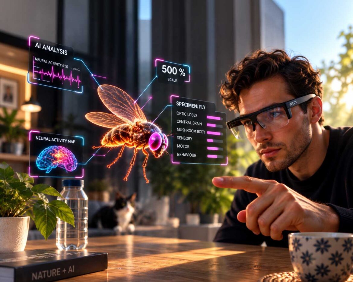
A hologram fly, driven by a real nervous system, reacts live to a real room.
Image: © Ulrich Buckenlei | XR Stager Online Magazine | VISORIC GmbH
On September 3, 2026, research teams led by Janelia FlyEM published the complete wiring diagram of an entire nervous system, every single neuron and every connection between them documented. Just nine days later, developers Pavlo Tkachenko and Stijn Spanhove showed what such a dataset is actually capable of once you let it run: they connected the nervous system to a pair of augmented reality glasses and brought a hologram to life that moved independently through a real living room, flying toward flowers and dodging a hand.
What looks at first glance like a quirky tech experiment stands in for a development that reaches far beyond one example: biological datasets, from neural maps to patient models to animal behavior, are increasingly being connected to artificial intelligence and spatial sensing, turning them from silent records into actively reacting systems.
- Two developers had an AI-simulated, real brain act independently in both digital and real space.
- The foundation is a complete nervous system captured via electron microscopy.
- The market for digital twins in healthcare is growing at double- to triple-digit rates per year.
- Livestock farming and conservation already use AI-driven, reacting behavior models too.
- The decisive principle is always the same: data plus model plus real environment.
This article breaks down how such a biological model is actually built, why a dataset alone still produces no behavior, where this idea already shows up in medicine, agriculture, and conservation, and where its limits lie.
The Moment a Dataset Turns Into Behavior
Every company and every research institution sits on datasets that describe something but do nothing. A process manual describes a workflow but doesn’t react when that workflow changes. A patient record shows what happened, not what is happening right now. That kind of data is valuable and yet strangely lifeless, as long as nobody connects it to the real world.
On September 3, 2026, Janelia FlyEM, the University of Cambridge, the MRC Laboratory of Molecular Biology, and Google Research published the so-called MaleCNS connectome in the journal Cell, the complete wiring diagram of a male fruit fly’s nervous system, brain and nerve cord together: 166,700 neurons, 25.6 million documented connections, reconstructed from real electron microscope images.[1] Just days later, developers Pavlo Tkachenko and Stijn Spanhove connected this dataset to Snap Spectacles, a see-through pair of AR glasses, and let it turn into a hologram that reacted independently to a real living room, flying toward flowers, dodging an approaching hand, and landing on a real teacup.[2]
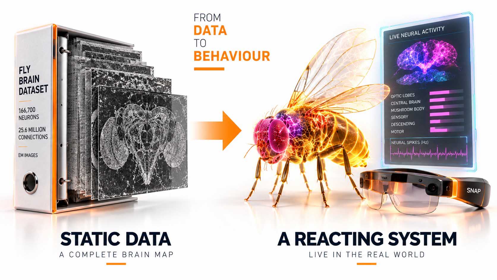
A silent wiring diagram turned into a reacting hologram within days.
Graphic: From passive dataset to active system | © Ulrich Buckenlei | XR Stager Online Magazine | VISORIC GmbH
That exact shift, from pure description to real, visible reaction, is the throughline of this article. As the following chapters show, it applies to far more than a single insect.
- The MaleCNS connectome maps 166,700 neurons and 25.6 million connections.
- Published on September 3, 2026 in the journal Cell.
- Two developers connected the dataset to AR glasses just days later.
- The result reacted live and unpredictably to a real room.
- A dormant 3D dataset became an active, observable system.
How such a biological dataset is actually built, and what opportunities that construction process opens up for biology itself, is the subject of the next chapter.
How Such a Model Is Built, and the Opportunity It Creates for Biology
A biological 3D dataset of this depth isn’t created on a drawing board but through the painstaking reconstruction of real tissue, and depending on scale, very different methods come into play.
For the MaleCNS connectome, real nerve tissue was sliced into thousands of ultra-thin layers, each layer photographed under an electron microscope, and every single nerve fiber then traced across all layers, both manually and automatically, a process needed for cell- and synapse-level resolution but enormously computation-heavy.[3] For larger biological structures, whole organs or organisms, other 3D reconstruction methods take over instead, such as CT and MRI imaging or photogrammetric and volumetric scanning, depending on whether the focus is cell-level detail or the entire body.
For such a wiring diagram to actually produce behavior, an additional computational model is needed. In the connectome’s case, that was a model published by researcher Philip Shiu and colleagues in Nature back in 2024, one already able to predict real behavior such as feeding or grooming.[4] For sufficient processing speed, the core was ported to specialized hardware, close enough to real time to feel like a living reaction rather than a sluggish simulation.
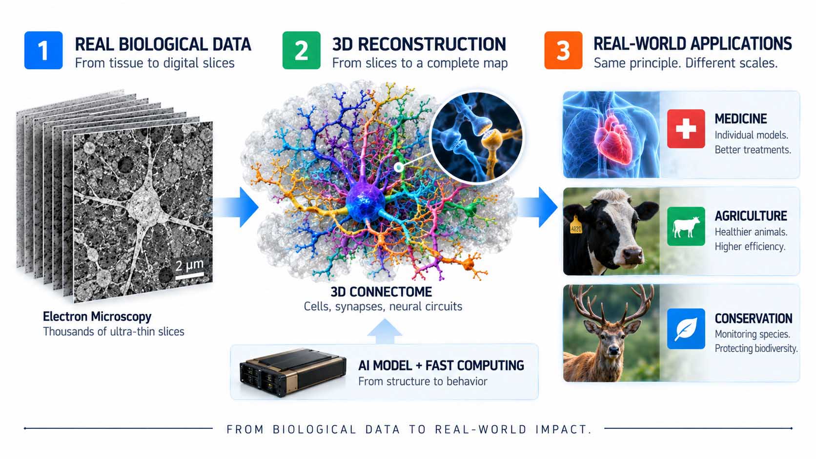
From tissue slice to a running map, and from there into three entirely different fields.
Graphic: Reconstruction methods and application fields for biological 3D datasets | © Ulrich Buckenlei | XR Stager Online Magazine | VISORIC GmbH
This construction process opens up an independent field of application for biology itself, separate from industry or technology. An individual health model, a livestock behavior model, or a model of an endangered species all follow the same basic principle: reconstruction first, then a model that derives behavior from it. This also opens new possibilities for research itself, hypotheses about behavior can be tested on a working digital model instead of every trial requiring a live animal.
- Electron microscopy reconstructs biological structures at the cellular level.
- CT, MRI, and photogrammetric scanning capture larger organs and organisms.
- An additional computational model is what first turns the map into actual behavior.
- Specialized hardware makes the simulation nearly real-time capable.
- The same construction principle applies equally to medicine, livestock farming, and conservation.
For such a model to actually produce behavior, the data foundation alone still isn’t enough. Why that is the case is the subject of the next chapter.
Why Data Alone Still Isn’t Behavior
A dataset, no matter how detailed, only describes relationships at first. It shows what’s connected to what, the way an org chart shows who reports to whom, without a single decision ever being made by that alone.
For such a description to turn into actual behavior, an additional model is needed, one that determines when a relationship becomes a concrete reaction. In the connectome example, certain biologically known “descending” neurons took on exactly this role, controlling turns, escape reflexes, walking backward, feeding, or grooming, with their activity translated directly into movement.[5] For a company, the principle is identical: a knowledge base or a process model alone makes no decisions. Only an additional rule-based or AI model working on top of that foundation turns it into something that actually reacts.
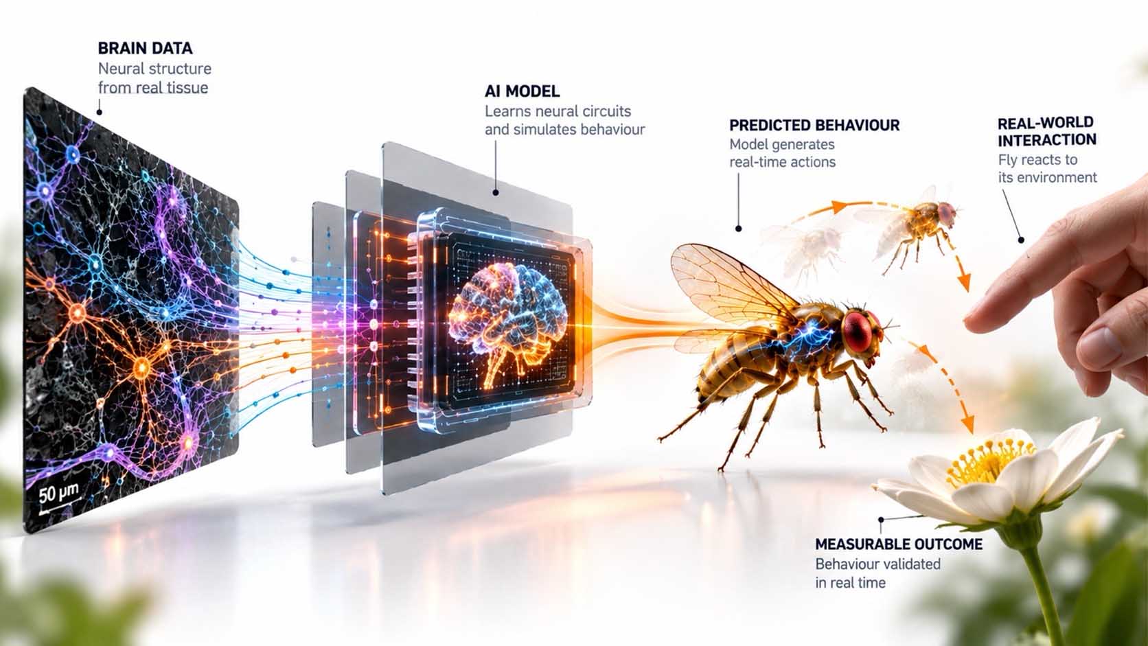
Between a data foundation and an actual action, there is always an additional decision model.
Graphic: From network diagram to concrete decision | © Ulrich Buckenlei | XR Stager Online Magazine | VISORIC GmbH
This distinction isn’t an academic nuance, it’s the exact point where many digitalization projects actually fail: building a database is the easier half of the work, making it truly capable of decisions is the harder one.
- A dataset shows only relationships, not decisions.
- In the connectome, certain neurons translate activity directly into movement.
- Companies likewise need an additional decision model layered on top of the data foundation.
- Building the data foundation is usually the easier part of the work.
- Without a decision layer, every dataset remains inconsequential.
Even with a decision model, a system remains inconsequential as long as it doesn’t know what’s actually happening around it right now. That missing connection is the subject of the next chapter.
The Missing Piece: A Connection to the Real Environment
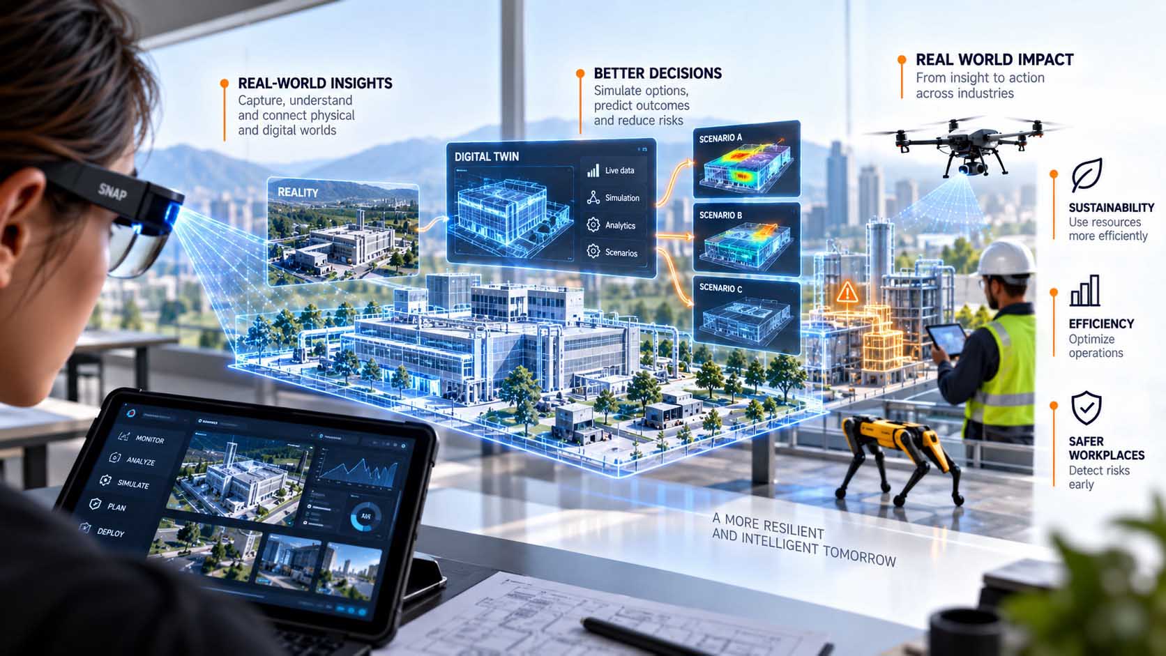
Even a dataset paired with a decision model remains inconsequential as long as it doesn’t know what’s actually happening around it right now. This is exactly where spatial computing comes in: it supplies the ongoing, up-to-date information about the real environment that a model needs to react correctly in the moment rather than just in theory.
In the connectome project, a pair of AR glasses took on this role. Snap Spectacles already have an understanding of the surrounding room, surfaces, depth, objects, hand movements, which the developers fed directly into the simulated nervous system.[6] For a company, that same role could just as well be a camera on a production floor, a sensor on a machine, or a wearable on a patient. In medicine, this is already happening at scale: the market for digital twins in healthcare is estimated at roughly 7.5 billion US dollars in 2026 and is expected to grow to over 101 billion US dollars by 2031, driven precisely by this ongoing connection between patient models and real, continuously captured measurement data.[7]
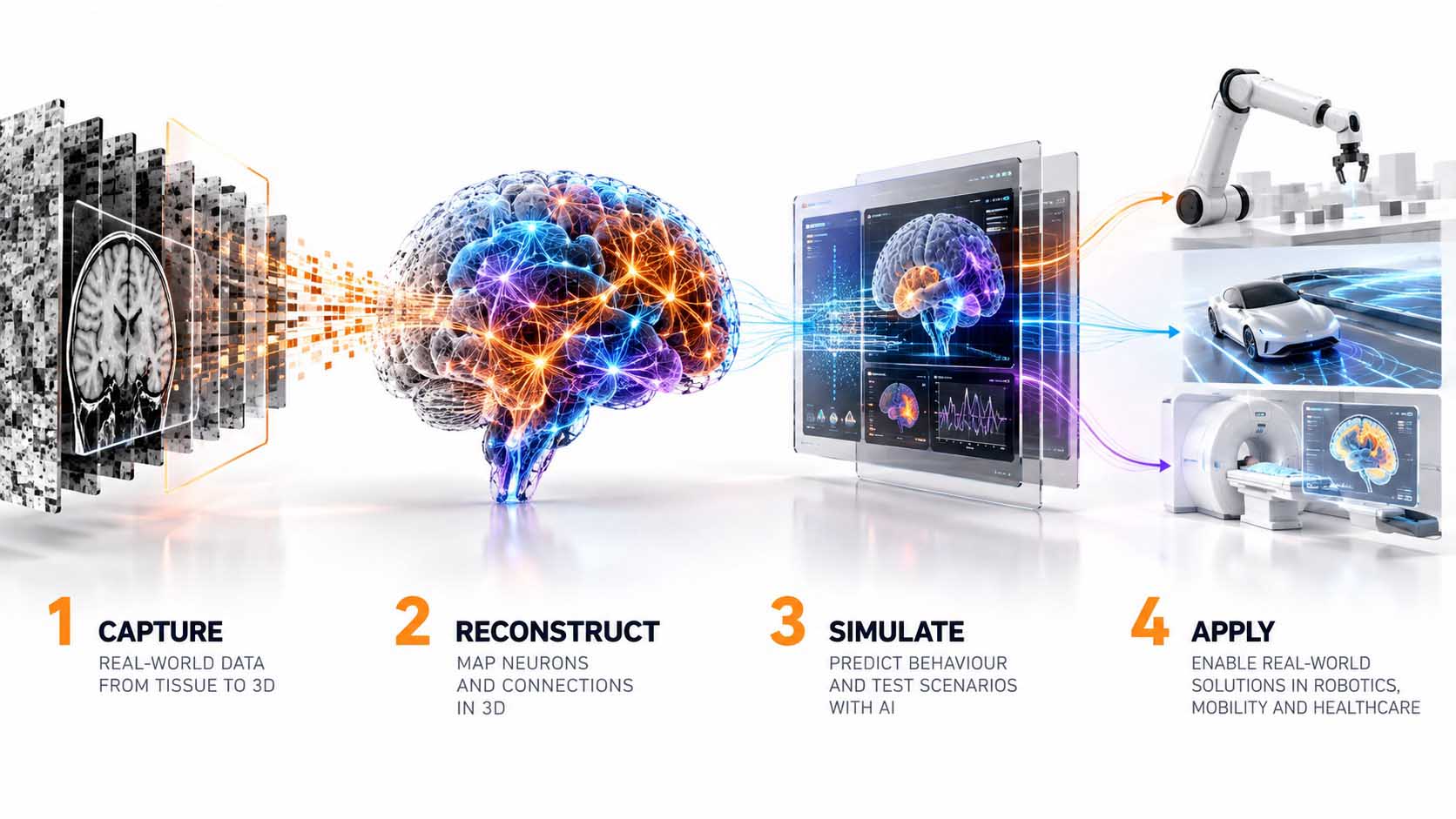
Whether AR glasses, a factory sensor, or a patient wearable, what matters is the ongoing connection to reality.
Image: Spatial computing as a sensor interface for decision models | © Ulrich Buckenlei | XR Stager Online Magazine | VISORIC GmbH
What matters here isn’t the specific technology, but that a model only becomes practically applicable, rather than purely theoretical, through this kind of ongoing connection to reality.
- Snap Spectacles gave the connectome project an understanding of the real room.
- The healthcare digital twin market grows from 7.5 to over 101 billion US dollars between 2026 and 2031.
- This growth is directly based on connecting patient models to real measurement data.
- The specific sensor technology is interchangeable, AR glasses, camera, or wearable.
- Without an ongoing connection to reality, every model remains purely theoretical.
Raw sensor data, however, is rarely directly usable for a decision model. How raw readings become understandable signals is the subject of the next chapter.
Translating Raw Data Into Understandable Signals
A camera delivers pixels, not the knowledge that a person is approaching or that a reading has become critical. That’s why a translation step is needed: an additional AI that turns raw sensor data into the kind of information the actual decision model can work with.
In the connectome project, Google’s Gemini AI model took on this task, recognizing objects in the room, a cup, a vase, a cat, and translating them, along with the glasses’ distance measurements, into the kind of sensory signal the simulated nervous system actually expects.[8] Fast movements nearby were interpreted as danger, the same stimulus that makes a real animal flinch. The same translation layer shows up in conservation research: AI models such as MegaDetector, developed at the California Institute of Technology, or the SpeciesNet model built on top of it, automatically search camera-trap images, detect and count animal species, and turn millions of raw images into usable population data, without any manual review.[9]
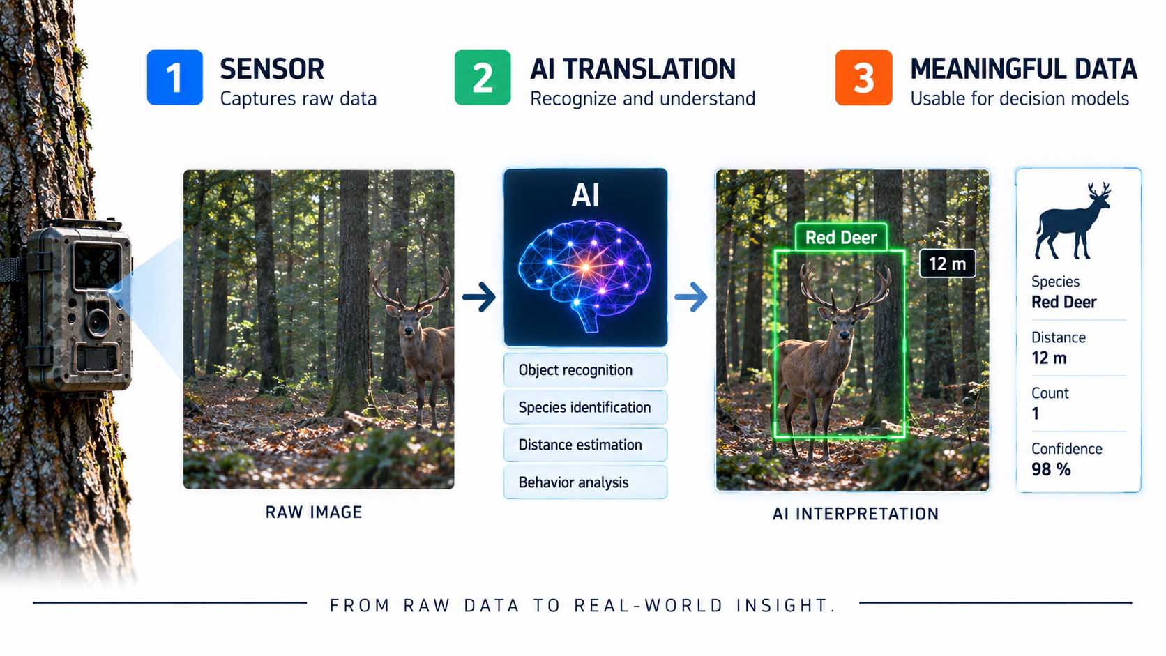
Only translation turns raw images into usable, species-accurate information.
Graphic: AI-driven object recognition as a translation layer between sensor and model | © Ulrich Buckenlei | XR Stager Online Magazine | VISORIC GmbH
Whether it’s a fly’s brain or a conservation camera, without this intermediate step even the best sensor data remains meaningless to a decision model.
- Gemini translated recognized objects into sensory signals in the connectome project.
- Fast movements were specifically interpreted as danger.
- MegaDetector and SpeciesNet take on the same translation role in conservation research.
- Both systems turn millions of camera-trap images into usable species data.
- Without an AI translation layer, raw sensor data remains useless to a model.
Only once all these building blocks come together does something emerge that looks, to an outside observer, like an independent, visible reaction. That’s exactly what the next chapter shows.
When a System Starts to React Visibly
Only once dataset, decision model, spatial sensing, and translated raw data come together does something emerge that looks like an independent reaction. In the connectome project, the hologram dodged an approaching hand, moved toward a real flower, and paused hesitantly on a teacup, exactly as the underlying “stop” neuron would predict, visible and comprehensible to anyone watching the scene.[10]
For companies and research institutions alike, this exact moment, when a system reacts visibly and comprehensibly, is the actual point of value creation. A health alert that appears the instant a problem arises, instead of in the next report, or a behavioral anomaly in livestock flagged in real time instead of at the next routine check, follows the same principle: data, model, and real environment visibly interlock.
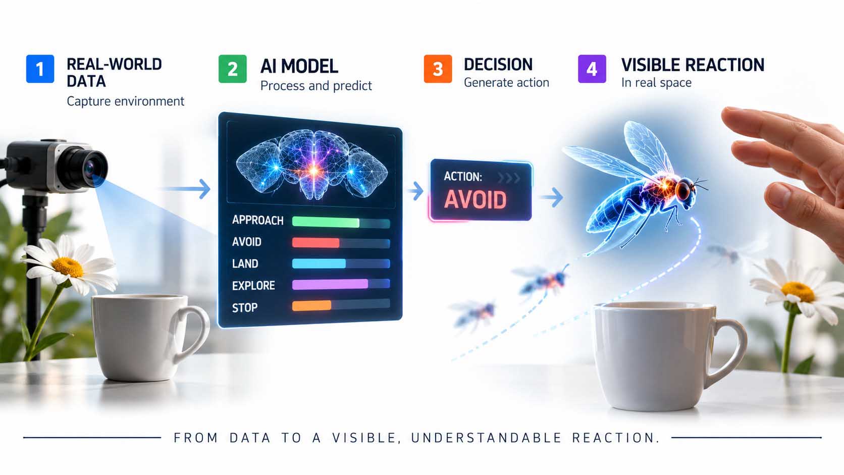
The moment a model reacts visibly is the actual point of value creation.
Graphic: From decision signal to a visible, comprehensible reaction | © Ulrich Buckenlei | XR Stager Online Magazine | VISORIC GmbH
As the next chapter shows in detail, this principle can already be demonstrated concretely across several real biological application fields today.
- The hologram reacted comprehensibly to real objects and movement in the video.
- Visible, situational reaction is a system’s actual point of value creation.
- An alert at the moment of the problem replaces the after-the-fact report.
- The same principle applies to health data as it does to animal behavior data.
- Data, model, and environment must visibly interlock.
How concretely this principle is already being applied today in medicine, agriculture, and conservation is shown by the following chapter, drawing on real projects and market figures.
Digitizing Living Organisms: Three Real-World Application Fields
Digitizing living organisms isn’t a side note to the principle described here, it’s already one of its most concrete application fields today, with solid projects and growing market figures across at least three areas.
In medicine, Dassault Systèmes’ Living Heart Project has been running since 2014, a detailed virtual model of the human heart that simulates electrical, structural, and fluid-dynamic processes. Together with the US Food and Drug Administration (FDA), this produced the so-called Enrichment Playbook in 2024, an officially recognized guide for how virtual heart simulations can serve as digital evidence in the approval of new cardiac devices, in some cases replacing animal testing.[11] 2025 brought an AI-powered, fully parametric version of the model that adapts to individual patient anatomy.[12]
In agriculture, the same principle is already widely deployed under the term precision livestock farming: sensors, ear tags, and camera systems from providers such as Afimilk, DeLaval, or Cainthus monitor livestock behavior, health, and fertility cycles in real time and flag anomalies before a veterinarian would spot them at the next routine check. The global market for AI in precision livestock farming grew to roughly 2.7 billion US dollars in 2025 and is expected to exceed 8 billion US dollars by 2030.[13]
In conservation, AI models such as MegaDetector and SpeciesNet handle the automated analysis of camera-trap images in protected areas worldwide, complemented by bioacoustic sensor networks that identify endangered bird species or nocturnal wildlife movement by sound, often running directly on low-power hardware on site, with no continuous internet connection required.[14]
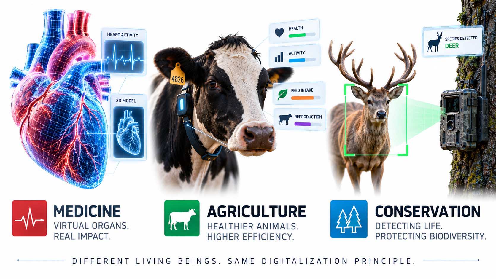
Three entirely different living organisms, one and the same digitization principle.
Infographic: Real-world application fields of biological digitization in medicine, agriculture, and conservation | Graphic: © Ulrich Buckenlei | XR Stager Online Magazine | VISORIC GmbH
| Application Field | Example Project | Market Size / Figure |
|---|---|---|
| Medicine | Living Heart Project (Dassault Systèmes, FDA) | Healthcare digital twin market: 7.5 to over 101 bn USD (2026–2031) |
| Agriculture | Precision Livestock Farming (Afimilk, DeLaval, Cainthus) | 2.7 to over 8 bn USD (2025–2030) |
| Conservation | MegaDetector, SpeciesNet, bioacoustic monitoring | Worldwide deployment across camera-trap networks, no single unified market value |
Three industries, one shared principle, reacting rather than purely descriptive datasets.
Table: Real-world application fields of biological digitization, as of summer 2026 | Source: manufacturer data, FDA, market research reports | © Ulrich Buckenlei | XR Stager Online Magazine | VISORIC GmbH
These three fields show that the principle described at the outset, dataset, model, and real environment, has long since moved beyond research labs into productive, commercial applications.
- The Living Heart Project has officially served as digital evidence for FDA approval since 2024.
- The healthcare digital twin market is growing at double- to quadruple-digit percentage rates.
- Precision livestock farming grows from 2.7 to over 8 billion US dollars by 2030.
- MegaDetector and SpeciesNet automate conservation monitoring worldwide.
- All three fields follow the same basic principle as the connectome example.
As impressive as these examples are, none of them work without careful technical and professional boundaries. What such systems can’t do on their own is the subject of the final chapter.
What Such a System Can’t Do on Its Own
As impressive as the results shown may be, they don’t replace careful technical and professional development. A decision model alone knows no physical constraints, no safety requirements, and no medical or operational conditions.
The developers of the connectome project state this openly themselves: the simulated nervous system ran on an ordinary laptop, not inside the glasses themselves, and things like leg rhythm, solid walls, or the precise physics of landing had to be added separately by hand, because a brain alone knows no physics.[15] A comparable limit applies in the medical context too: even the Living Heart model, developed over more than a decade, explicitly serves the FDA as supplementary, not sole, evidence, human review and classical clinical data remain part of the approval process.[16]
The assessment can come from data, reliable implementation remains handcrafted work.
Graphic: Limits of automated biological digitization | © Ulrich Buckenlei | XR Stager Online Magazine | VISORIC GmbH
For companies and biological applications alike, the same rule applies: the assessment of what would be right in a given situation can increasingly come from data and models, the reliable, safe, and compliant implementation of that assessment in the physical world remains the task of careful technical and professional development.
- A decision model knows no physics or safety requirements of its own.
- In the connectome project, movement physics and solid obstacles had to be added separately.
- Even the Living Heart model serves the FDA only as supplementary, not sole, evidence.
- Human review remains central in medicine, agriculture, and conservation.
- Assessment can come from data, reliable implementation remains handcrafted work.
Just how convincing the principle described at the outset already is in practice is shown most vividly in the following video.
Watching a Real Nervous System React, Live
The chapters above have shown how a biological 3D dataset is built, why it produces no behavior on its own, what role spatial sensing and AI translation play, and where this principle is already being applied today in medicine, agriculture, and conservation. It’s most vivid in the project that opened this article.
Embedded here is the video of the connectome project by Pavlo Tkachenko and Stijn Spanhove, showing a hologram fly being steered live through a real room, including a second screen displaying the neuron activity of the underlying model.[17]
Video: FlyBrain Spectacles, live-driven hologram based on the MaleCNS connectome | Project: Pavlo Tkachenko and Stijn Spanhove (pavlo-stijn.dev), reposted via Instagram @canmatrixx | © Ulrich Buckenlei | XR Stager Online Magazine | VISORIC GmbH
The video makes visible what was broken down technically in the previous chapters: a real dataset, a decision model, spatial sensing, and an AI translation layer interlock so precisely that an observer sees an independent, comprehensible reaction, not by chance, but as the result of the same structure that also underlies heart simulations, barn sensors, and conservation cameras.
- Video shows the real connectome project by Tkachenko and Spanhove.
- A second screen makes the model’s decision-making visible.
- The same underlying structure also carries medical, agricultural, and ecological applications.
- The technology becomes invisible exactly when all the building blocks fit together.
- This principle can be transferred to many other biological application fields.
This example makes tangible where the digitization of biological systems already stands today, from a single research demo to a principle backing real, multi-billion-dollar markets.
From the Biological Idea to Your Own Digital Twin Project
A solid decision to launch your own digital twin project, whether in medical technology, agriculture, or industrial production, doesn’t come from a single impressive example, it comes from the same interplay this article has described: a clean dataset, a suitable decision model, and an ongoing connection to the real environment.
The expert team at VISORIC GmbH in Munich combines over 15 years of experience in 3D visualization, AI, and spatial computing with hands-on experience building reactive, spatially aware systems, exactly the building blocks that also matter when digitizing biological or industrial datasets.[18] VISORIC helps companies turn existing datasets, whether CAD models, sensor logs, or training materials, into reactive, interactive systems instead of leaving them as pure archives.

15 years of experience in 3D, AI, and spatial computing, the VISORIC expert team from Munich.
Image: © Ulrich Buckenlei | XR Stager Online Magazine | VISORIC GmbH
A well-thought-out pilot project, a single dataset, a single training environment, can often be realized much faster and more cost-effectively than many companies expect. VISORIC guides this path from the initial assessment through visualizing possible application scenarios to a decision-ready proposal.[19]
- Assessment of whether and how an existing dataset can be digitally embodied.
- 3D visualization and spatial computing implementation of reactive systems.
- From the first pilot idea to a solid decision-ready proposal.
That’s exactly where the starting point for a conversation lies: not with the grand vision of a complete digital twin, but with a clearly defined, quickly implementable first step.
Want to find out whether and how your existing datasets can be turned into a reactive, spatially aware system?
Talk to the VISORIC expert team in Munich about feasibility, model choice, and visualizing possible application scenarios.
Contact:
Email: info@visoric.com
Phone: +49 89 21552678
Sources and References
- MaleCNS connectome research publication, Cell (September 3, 2026); Janelia FlyEM MaleCNS project page (male-cns.janelia.org).
- Blog post pavlo-stijn.dev/blog/posts/a-real-fly-brain-on-spectacles.html (September 12, 2026), Pavlo Tkachenko and Stijn Spanhove.
- Janelia FlyEM MaleCNS download page; GitHub github.com/philshiu/Drosophila_brain_model.
- Shiu et al., Nature (2024); GitHub github.com/PtPavloTkachenko/fly-brain-spectacles.
- Blog post pavlo-stijn.dev, section on the function of descending neurons.
- Blog post pavlo-stijn.dev; Instagram repost via @canmatrixx (video evidence).
- Digital Twins in Healthcare Global Market Report 2026, ResearchAndMarkets / GlobeNewswire, August 2026.
- Blog post pavlo-stijn.dev, section on Gemini object recognition; GitHub repository fly-brain-spectacles.
- Beery, S., The MegaDetector, California Institute of Technology (2023); PubMed, Fine-tuning SpeciesNet for wildlife classification (2026).
- Instagram video via @canmatrixx; blog post pavlo-stijn.dev.
- Dassault Systèmes, Living Heart Project and Enrichment Playbook, FDA collaboration (2024).
- Dassault Systèmes, press release on AI-powered virtual twins of the Living Heart Project (2025).
- The Business Research Company, AI In Precision Livestock Farming Market Size Report 2026-2030.
- Beery, S., The MegaDetector (Caltech, 2023); Frontiers in Conservation Science, Adaptive AI for wildlife monitoring (2026).
- Blog post pavlo-stijn.dev, “honest note” section.
- Research & Development World, How the Living Heart Project could transform FDA’s approach to device testing (2024).
- Blog post pavlo-stijn.dev; Instagram repost via @canmatrixx.
- VISORIC practice projects in 3D visualization, digital twins, and spatial computing.
- XR Stager platform for real-time 3D, digital twins, and industrial spatial computing applications.
Contact Persons:
Ulrich Buckenlei (Creative Director)
Mobile: +49 152 53532871
Email: ulrich.buckenlei@visoric.com
Nataliya Daniltseva (Project Manager)
Mobile: +49 176 72805705
Email: nataliya.daniltseva@visoric.com
Address:
VISORIC GmbH
Bayerstraße 13
D-80335 Munich